What Is a Ventral Hernia?
A hernia occurs when there is a hole in the muscles of the abdominal wall, allowing a loop of intestine or abdominal tissue to push through the muscle layer. A ventral hernia is a hernia that occurs at any location along the midline (vertical center) of the abdomen wall.

Types of Ventral Hernia
- Epigastric (stomach area) hernia: Occurs anywhere from just below the breastbone to the navel (belly button). This type of hernia is seen in both men and women.
- Umbilical (belly button) hernia: Occurs in the area of the belly button.
- Incisional hernia. Develops at the site of a previous surgery. Up to one-third of patients who have had an abdominal surgery will develop an incisional hernia at the site of their scar. This type of hernia can occur anytime from months to years after an abdominal surgery.
What Are The Causes for Developing a Ventral Hernia?
There are many causes including:
- Weakness at the incision site of a previous abdominal surgery (which could result from an infection at the surgery site or failed surgical repair/mesh placement).
- Weakness in an area of the abdominal wall that was present at birth.
- Weakness in the abdominal wall caused by conditions that put strain on the wall
- Frequent coughing episodes
- Severe vomiting
- Pregnancy
- History of lifting or pushing heavy objects
- Injuries to the bowel area
What Are The Signs and Symptoms of a Ventral Hernia?
Some patients don’t feel any discomfort in the early stages of ventral hernia formation. Often, the first sign is a visible bulge under the skin in the abdomen or an area that is tender to the touch. The bulge may flatten when lying down or pushing against it.
- Lifts heavy objects.
- Strains to have a bowel movement/urinate.
- Sits or stands for long periods of time.
How is a Ventral Hernia Diagnosed?
Your doctor will review your medical and surgical history. He or she will also perform a physical exam of the abdominal area where a ventral hernia may have occurred. Your doctor may then order imaging tests of the abdomen to look for signs of a ventral hernia. These tests may include an ultrasound, computed tomography (CT) scan or a magnetic resonance imaging (MRI) study.
How is a Ventral Hernia Repaired?
- Open hernia repair: An open incision is made in the abdomen where the hernia has occurred, and the intestine or abdominal tissue is pushed back into place. A mesh material is placed to reinforce this repair and reduce hernia recurrences. The skin is usually closed with dissolvable stitches and glue.
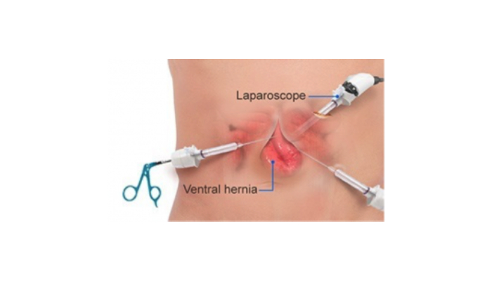
- Laparoscopic surgery: Several small incisions are made away from where the hernia has occurred. A laparoscope (a thin lighted tube with a camera on the tip) is inserted through one of the openings to help guide the surgery. A surgical mesh material may be inserted to strengthen the weakened area in the abdominal wall. Advantages of this approach compared with open hernia repair include a lower risk of infection, because smaller-sized incisions are used.
- Robotic hernia repair: Like laparoscopic surgery, robotic surgery uses a laparoscope, and is performed in the same manner (small incisions, a tiny camera). Robotic surgery differs from laparoscopic surgery in that the surgeon is seated at a console in the operating room, and handles the surgical instruments from the console. While robotic surgery can be used for some smaller hernias, or weak areas, it can now also be used to reconstruct the abdominal wall. Other benefits of robotic hernia surgery are that the patient has tiny scars rather than one large incision scar, and there may be less pain after this surgery compared to open surgery.
