Overview Of Esophagus
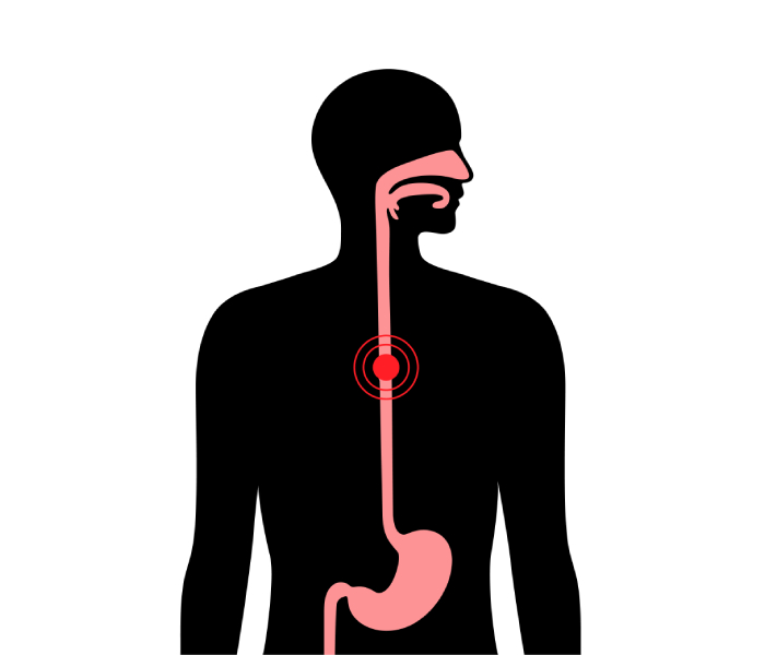
The esophagus is a muscular tube connecting the throat (pharynx) with the stomach. The esophagus is about 8 inches long, and is lined by moist pink tissue called mucosa. The esophagus runs behind the windpipe (trachea) and heart, and in front of the spine. Just before entering the stomach, the esophagus passes through the diaphragm.
The upper esophageal sphincter (UES) is a bundle of muscles at the top of the esophagus. The muscles of the UES are under conscious control, used when breathing, eating, belching, and vomiting. They keep food and secretions from going down the windpipe.
The lower esophageal sphincter (LES) is a bundle of muscles at the low end of the esophagus, where it meets the stomach. When the LES is closed, it prevents acid and stomach contents from traveling backwards from the stomach. The LES muscles are not under voluntary control.
Proper function of the esophagus and its sphincters is essential for healthy digestion and preventing conditions like acid reflux or GERD.

Esophagus Conditions
- Heartburn: An incompletely closed LES allows acidic stomach contents to back up (reflux) into the esophagus. Reflux can cause heartburn, cough or hoarseness, or no symptoms at all.
- Gastroesophageal reflux disease (GERD): When reflux occurs frequently or is bothersome, it’s called gastroesophageal reflux disease (GERD).
- Esophagitis: Inflammation of the esophagus. Esophagitis can be due to irritation (as from reflux or radiation treatment) or infection.
- Barrett’s esophagus: Regular reflux of stomach acid irritates the esophagus, which may cause the lower part to change its structure. Very infrequently, Barrett’s esophagus progresses to esophageal cancer.
- Esophageal ulcer: An erosion in an area of the lining of the esophagus. This is often caused by chronic reflux.
- Esophageal stricture: A narrowing of the esophagus. Chronic irritation from reflux is the usual cause of esophageal strictures.
- Achalasia: A rare disease in which the lower esophageal sphincter does not relax properly. Difficulty swallowing and regurgitation of food are symptoms.
- Esophageal cancer: Although serious, cancer of the esophagus is uncommon. Risk factors for esophageal cancer include smoking, heavy drinking, and chronic reflux.
- Mallory-Weiss tear: Vomiting or retching creates a tear in the lining of the esophagus. The esophagus bleeds into the stomach, often followed by vomiting blood.
Esophagus Tests
- Upper endoscopy, EGD (esophagogastroduodenoscopy): A flexible tube with a camera on its end (endoscope) is inserted through the mouth. The endoscope allows examination of the esophagus, stomach, and duodenum (small intestine).
- Esophageal pH monitoring: A probe that monitors acidity (pH) is introduced into the esophagus. Monitoring pH can help identify GERD and follow the response to treatment.
- Barium swallow: A person swallows a barium solution, then X-ray films are taken of the esophagus and stomach. Most often, a barium swallow is used to seek the cause of difficulty swallowing.
How are candidates for Liver Transplant Determined?Esophagus Treatments
- H2 blockers: Histamine stimulates acid release in the stomach. Certain antihistamines called H2 blockers can reduce acid, improving GERD and esophagitis.
- Proton pump inhibitors: These medicines turn off many of the acid-producing pumps in the stomach wall. Reduced stomach acid can reduce GERD symptoms, and help ulcers or esophagitis to heal.
- Esophagectomy: Surgical removal of the esophagus, usually for esophageal cancer.
- Esophageal dilation: A balloon is passed down the esophagus and inflated to dilate a stricture, web, or ring that interferes with swallowing.
- Esophageal variceal banding: During endoscopy, rubber band-like devices can be wrapped around esophageal varices. Banding causes varices to clot, reducing their chance of bleeding.
- Biopsy: Often done through an endoscope, a small piece of the esophagus is taken to be evaluated under a microscope.
- Confocal laser endomicroscopy: A new procedure that takes the microscope inside a patient, which may replace the need for many biopsies.
Schedule Your Consultation Today!
For expert care related to esophageal health, acid reflux, GERD, or other digestive concerns, consult Dr. Ujwal Zambare, the best Gastroenterologist and GI Surgeon in Wakad, Pune. Get an accurate diagnosis and personalized treatment plan to ensure optimal digestive health.
